Bring AI to your
medical imaging research
From scan to study results in your browser: segment with AI, measure every structure, and compare patient groups.
- AI models for CT and MRI
- No installation or coding required
- Click-to-segment tools & manual editing
- Quantitative CT, MRI, PET & SPECT analysis
- Export your results
Free to start. No credit card needed.
Users in more than 20 countries.


Segment with clicks. Guide the AI to outline anatomy on your CT or MRI scan.
Review your results. Refine boundaries with smart brushes and interpolation between slices.
Quantify PET/CT. Measure SUVmax, SUVmean, SUVpeak, volume, and more for each structure.
From upload to measurement in five steps
Upload your
medical scans
Pick an AI model
from our library
Run AI tasks
in the cloud
View, adapt, and
download results
Measure and compare
in
Measurements
Every segmented structure is measured in your browser.
- Volume
- Per structure
- Intensity
- Mean, median, and range
- PET and SPECT
- Functional values, such as SUV
- Tables and reports
- A table per patient, a report per study
- Trends
- Across each patient's scan dates
- Compare groups
- Patient groups you define
Segment · Report · Analyse - One platform
Interactive and automatic segmentation
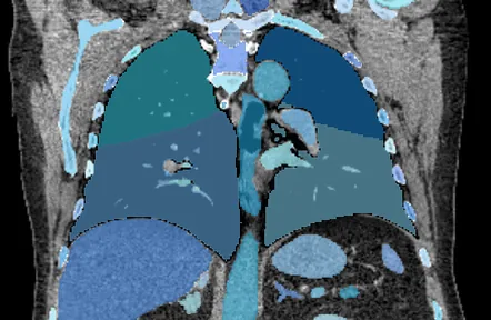
Segment organs, bones and muscles on CT and MRI scans, automatically or with interactive mouse clicks. Review and edit image segmentations in your browser.


Synthetic image generation
Generate synthetic CT or brain MRI images without an input scan, for research datasets, software testing, and demonstrations.
- Model
- MAISI-v2
- Task
- CT (rflow-ct)
- Mode
- image-mask
- Body region
- abdomen
- Dimensions
- 512 × 512 × 128 voxels
- Spacing
- 1.00 × 1.00 × 3.00 mm

- Model
- MAISI-v2
- Task
- MR brain (rflow-mr-brain)
- Mode
- image-only
- Contrast
- T2
- Dimensions
- 256 × 256 × 256 voxels
- Spacing
- 0.95 × 0.95 × 0.70 mm

Research reports from CT
Generate structured research reports from chest and abdominal CT scans.

2. Bilateral renal cysts. [findings list] Renal cyst
Abdominal vascular abnormality
Abdominal vascular calcification
Run your first AI task
Free to start. No credit card needed.