Heart chambers and great vessels on CT
MOOSE segments the four heart chambers and the myocardium on a CT scan, plus the aorta, pulmonary artery, inferior vena cava, portal and splenic vein, and left and right iliac arteries and veins. Studies use these outlines to measure chamber volumes, compare vessel size along the trunk, and relate both to health outcomes. On PET or SPECT, blood in the aorta or the heart chambers serves as a reference region, and uptake can be read in the myocardium and along the aorta.
Find answers to research questions like: How does myocardial uptake, relative to blood-pool uptake, change between scan dates? How closely do CT chamber volumes follow cardiac MRI values? How large is the pulmonary artery compared with the aorta?
Run it online in your browser, with no coding.
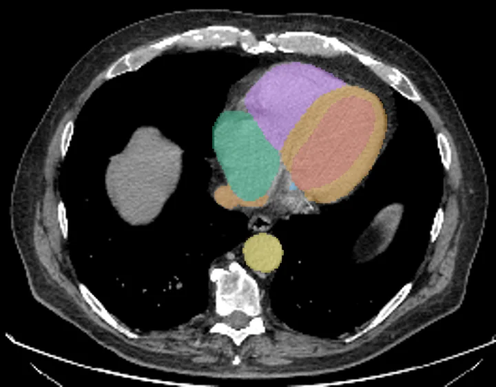
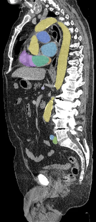
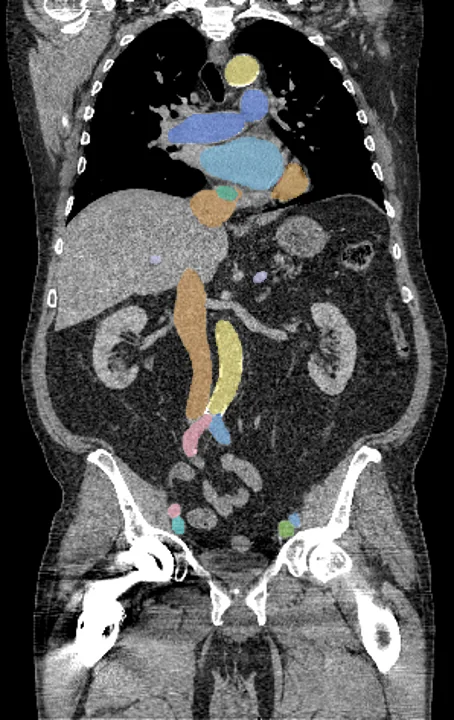
Found in this example
- Heart myocardium
- Heart atrium left
- Heart atrium right
- Heart ventricle left
- Heart ventricle right
- Aorta
- Iliac artery left
- Iliac artery right
- Iliac vena left
- Iliac vena right
- Inferior vena cava
- Portal splenic vein
- Pulmonary artery
Example scan: neck to thigh CT, TotalSegmentator dataset