Lung lobe segmentation on CT
Lung lobe segmentation on CT with MOOSE outlines both lungs by lobe: upper and lower on the left; upper, middle, and lower on the right. Lobe-by-lobe volumes let researchers build reference values, compare groups, and see how each lobe changes between breath-in and breath-out scans. On SPECT or PET, each lobe's counts or uptake show how function is spread across the lung.
Find answers to research questions like: How do lobe volumes vary with age, sex, and height? Which lobes change most between breath-in and breath-out? Does a lobe's share of total uptake change between scan dates?
Run this task →
Run it online in your browser, with no coding.
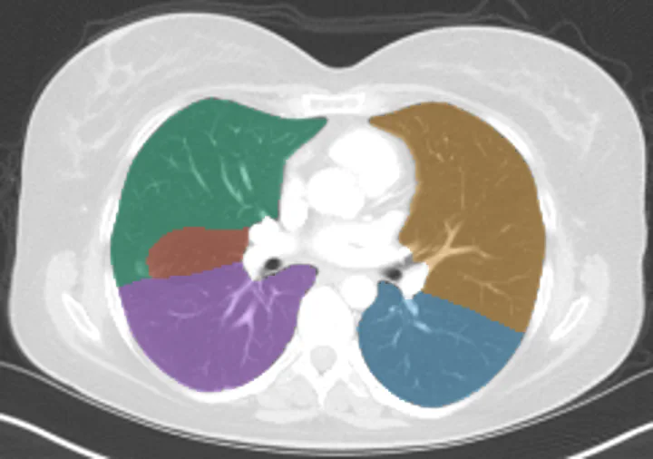
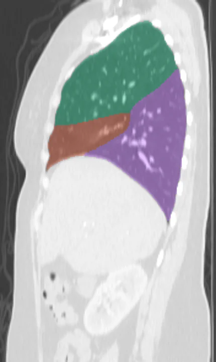
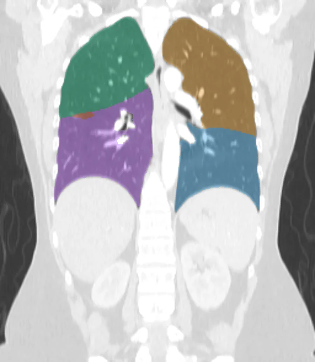
Found in this example
- Lung upper lobe left
- Lung lower lobe left
- Lung upper lobe right
- Lung middle lobe right
- Lung lower lobe right
Example scan: chest and upper abdomen CT, TotalSegmentator dataset