Organ segmentation on CT
MOOSE CT segmentation of the organs outlines 19 structures on a CT scan: brain, thyroid lobes, trachea, five lung lobes, liver, gallbladder, spleen, pancreas, stomach, adrenal glands, kidneys, and bladder, with left and right kept apart for paired organs. Research groups use it to measure organ volumes and Hounsfield units across a cohort. On PET or SPECT, it gives SUV per organ, which can be set against a reference organ or followed between scan dates.
Find answers to research questions like: How do liver, spleen, pancreas, and kidney volumes differ between groups? Which organs give the least consistent volumes across segmentation tools? Which organs link most strongly to each other in whole-body uptake?
Run it online in your browser, with no coding.
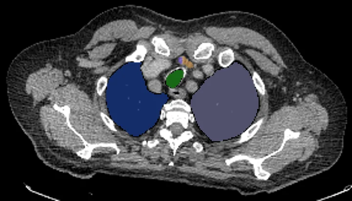
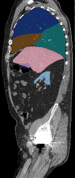
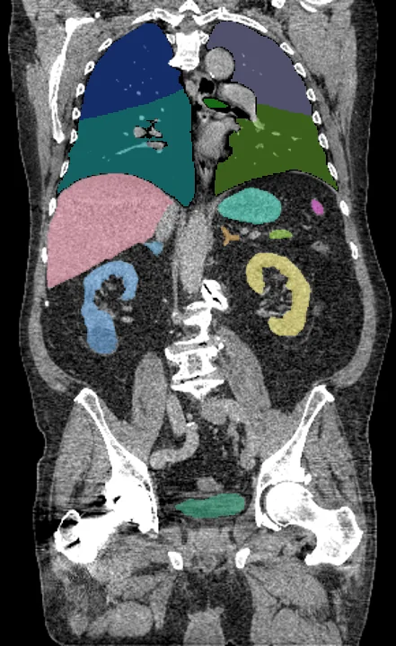
Found in this example
- Adrenal gland left
- Adrenal gland right
- Bladder
- Gallbladder
- Kidney left
- Kidney right
- Liver
- Lung lower lobe left
- Lung lower lobe right
- Lung middle lobe right
- Lung upper lobe left
- Lung upper lobe right
- Pancreas
- Spleen
- Stomach
- Thyroid left
- Thyroid right
- Trachea
Also finds: Brain
Example scan: neck to thigh CT, TotalSegmentator dataset