Peripheral bone segmentation on CT
The skeleton outside the spine, rib cage, and pelvis, as MOOSE outlines it on a CT scan: skull, clavicle, scapula, humerus, radius, ulna, carpals, metacarpals, fingers, femur, patella, tibia, fibula, tarsals, metatarsals, and toes, with left and right kept apart and only the bones in view outlined. Researchers use it to compare bone volumes between groups and sides, follow change over time, and assign findings to the bone they sit in. On PET or SPECT, uptake can be read bone by bone and compared between the two sides.
Find answers to research questions like: Do left and right limb bones differ in volume within one person? How does uptake in a hand or foot bone differ between the two sides of the body? How does the shape of a single hand or foot bone vary between people?
Run it online in your browser, with no coding.
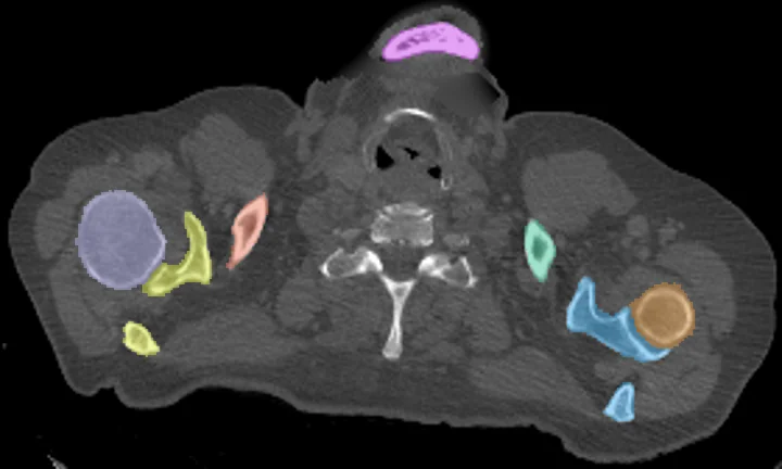
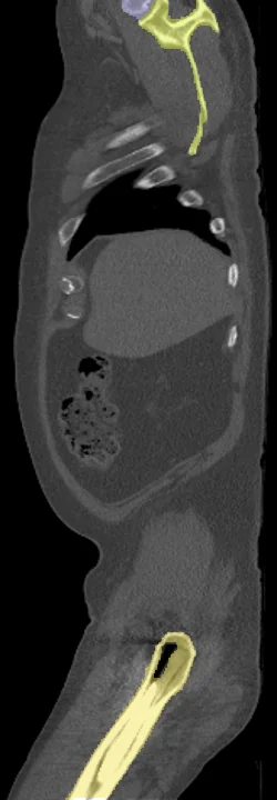
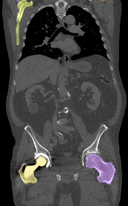
Found in this example
- Clavicle left
- Clavicle right
- Femur left
- Femur right
- Humerus left
- Humerus right
- Scapula left
- Scapula right
- Skull
Also finds: Carpal left, Carpal right, Fibula left, Fibula right, Fingers left, Fingers right, Metacarpal left, Metacarpal right, Metatarsal left, Metatarsal right, Patella left, Patella right, Radius left, Radius right, Tarsal left, Tarsal right, Tibia left, Tibia right, Toes left, Toes right, Ulna left, Ulna right
Example scan: neck to thigh CT, TotalSegmentator dataset