Vertebrae and pelvis segmentation on CT
Each vertebra from C1 down to L6 has its own outline in this spine segmentation on CT, and MOOSE also outlines the sacrum and the left and right hip bones. Studies use it to count vertebrae, find variants at the border of the lumbar spine and sacrum, and place muscle and fat measurements at the same level in every participant. On PET or SPECT, uptake can be read bone by bone across the spine, sacrum, and hip bones, set against a reference region such as the liver.
Find answers to research questions like: How common are extra or fused segments where the lumbar spine meets the sacrum? How do sacrum and hip bone dimensions differ between women and men? How does uptake differ between the spine, the sacrum, and the hip bones?
Run it online in your browser, with no coding.
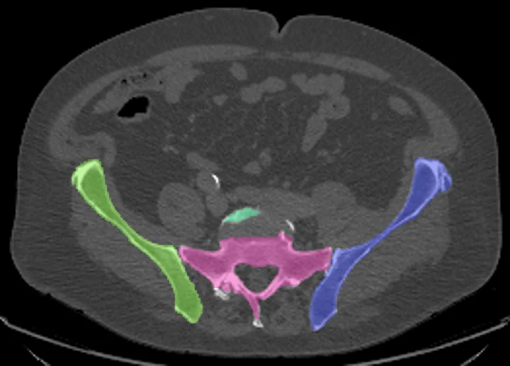
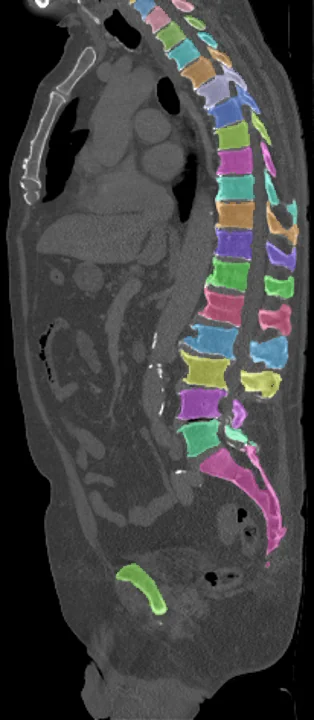
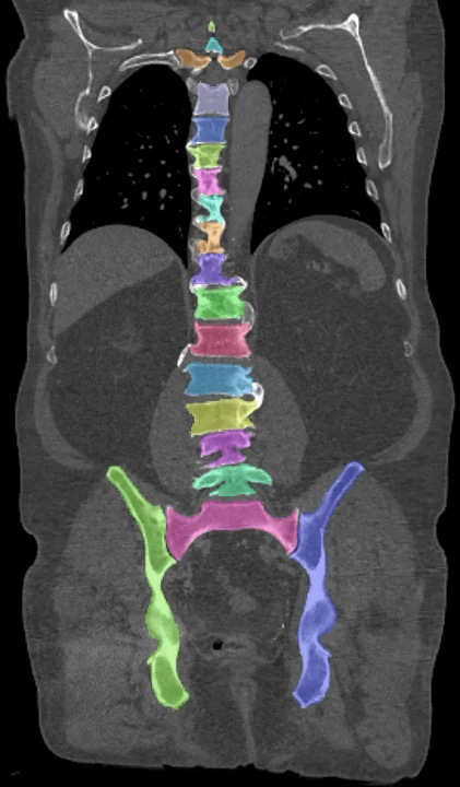
Found in this example
- Vertebra C6
- Vertebra C7
- Vertebra T1
- Vertebra T2
- Vertebra T3
- Vertebra T4
- Vertebra T5
- Vertebra T6
- Vertebra T7
- Vertebra T8
- Vertebra T9
- Vertebra T10
- Vertebra T11
- Vertebra T12
- Vertebra L1
- Vertebra L2
- Vertebra L3
- Vertebra L4
- Vertebra L5
- Hip left
- Hip right
- Sacrum
Also finds: Vertebra C1, Vertebra C2, Vertebra C3, Vertebra C4, Vertebra C5, Vertebra L6
Example scan: neck to thigh CT, TotalSegmentator dataset