Chest to hip anatomy on CT
CT segmentation with MRSegmentator outlines 40 structures between the chest and the hips, among them the liver, kidneys, bowel, heart, lungs, aorta, spine, femurs, and gluteus muscles. CT and MRI share one list of structures, so a study can set CT and MRI cohorts side by side and compare the same structure across both. On PET or SPECT, the same outlines read uptake in the organs, the spine, and the surrounding muscles.
Find answers to research questions like: Do CT and MRI give the same volume for the same structure in the same people? Do differences between them need correcting before cohorts are pooled? How does uptake in the abdominal organs differ between groups?
Run it online in your browser, with no coding.
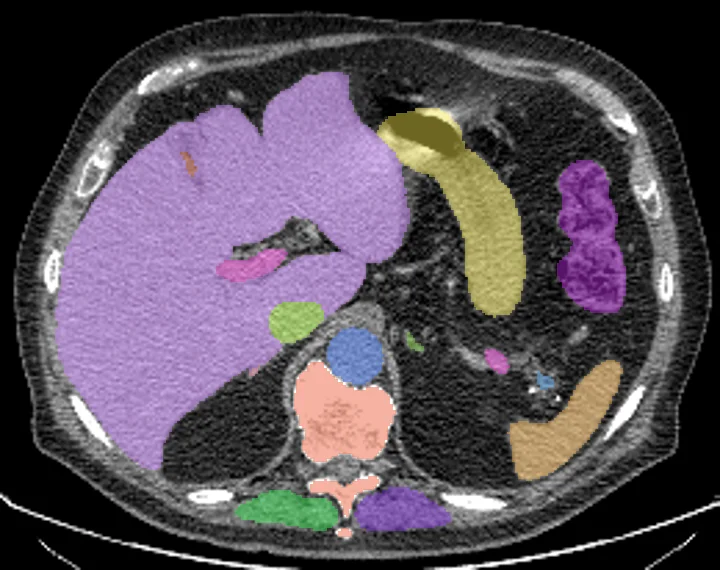
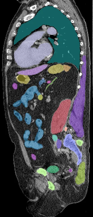
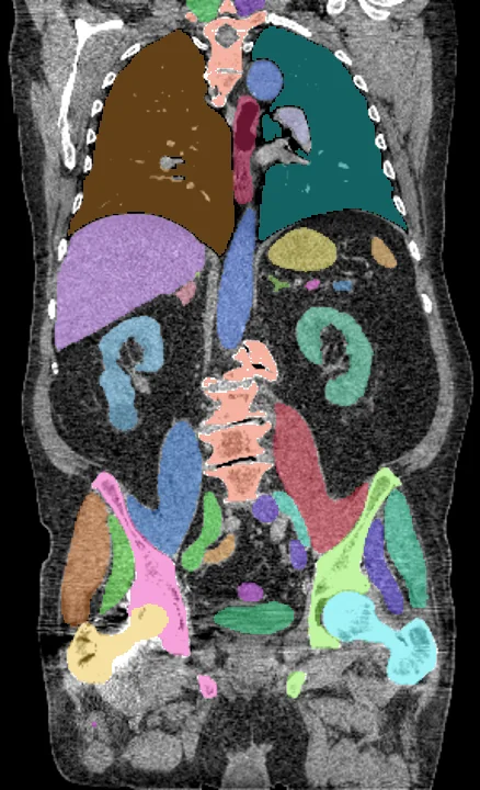
Found in this example
- Spleen
- Right kidney
- Left kidney
- Gallbladder
- Liver
- Stomach
- Pancreas
- Right adrenal gland
- Left adrenal gland
- Left lung
- Right lung
- Heart
- Aorta
- Inferior vena cava
- Portal vein and splenic vein
- Left iliac artery
- Right iliac artery
- Left iliac vena
- Right iliac vena
- Esophagus
- Small bowel
- Duodenum
- Colon
- Urinary bladder
- Spine
- Sacrum
- Left hip
- Right hip
- Left femur
- Right femur
- Left autochthonous muscle
- Right autochthonous muscle
- Left iliopsoas muscle
- Right iliopsoas muscle
- Left gluteus maximus
- Right gluteus maximus
- Left gluteus medius
- Right gluteus medius
- Left gluteus minimus
- Right gluteus minimus
Example scan: neck to thigh CT, TotalSegmentator dataset