Airway segmentation on CT
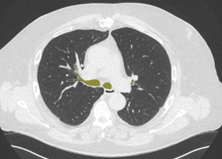
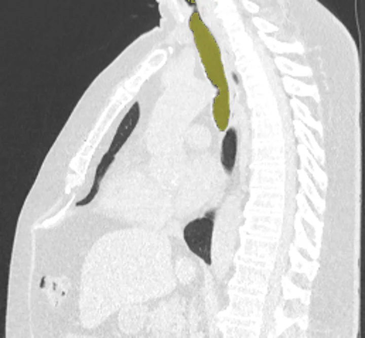
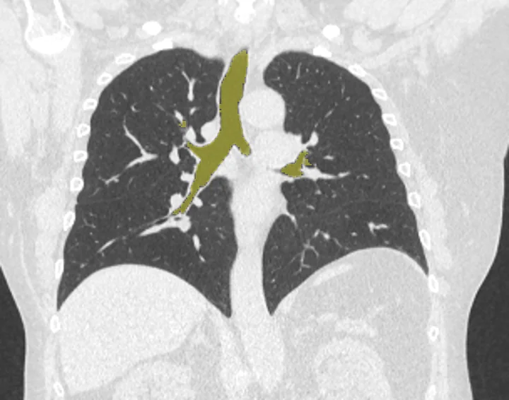
On CT scans that include the chest, NV-Segment-CT outlines the airway as one 3D structure. Each scan is read at 1.5 mm resolution, so the smallest branches come out coarse. Studies use this airway segmentation to measure airway tree size against lung size, compare breath-in and breath-out scans, and build models for airflow simulation. On PET or SPECT, uptake or ventilation counts can be read along the airway and set beside its size.
Find answers to research questions like: How much does airway tree shape vary between people? How does airway tree size relate to lung size? How do ventilation scan counts relate to airway size?
Run this task →
Run it online in your browser, with no coding.