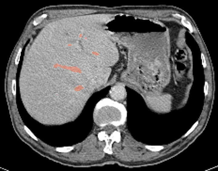
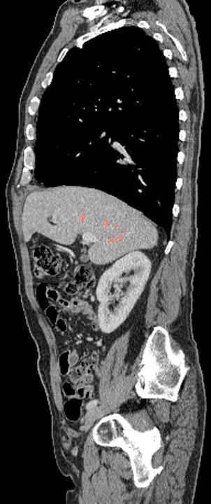
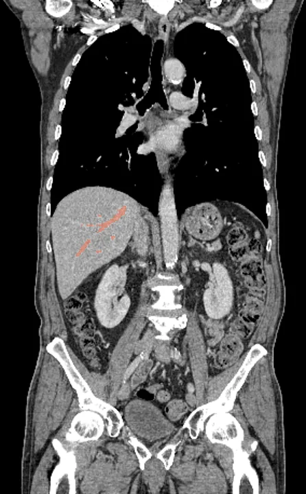
Liver vessel segmentation on CT
The blood vessels inside the liver come back from NV-Segment-CT as one 3D structure, named hepatic vessel. A contrast-enhanced CT scan in the portal venous phase is required: without contrast, little or nothing gets outlined. Studies use it to compare vessel volume and its share of the liver between groups, and to check contrast enhancement across scans. On PET or SPECT, the outline reads blood-pool uptake inside the vessels of the liver, which can be set against the liver tissue around them.
Find answers to research questions like: How do hepatic vessel volumes differ between groups? Does blood-pool uptake in the liver vessels stay steady between scan dates? How much do vessel Hounsfield values vary between scanners and contrast protocols?
Run it online in your browser, with no coding.