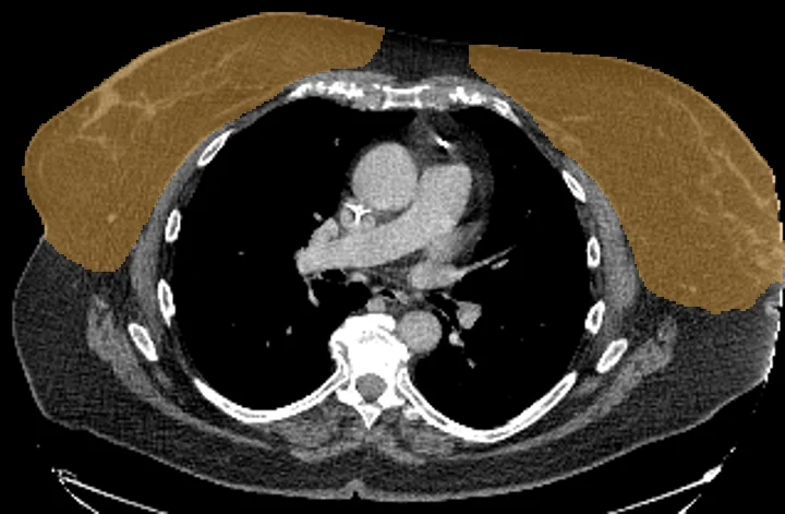
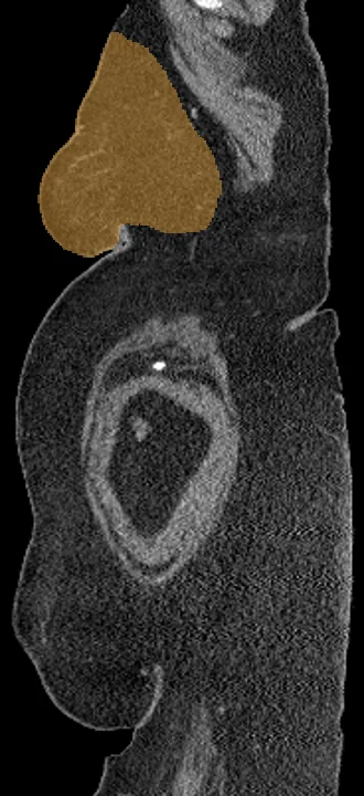
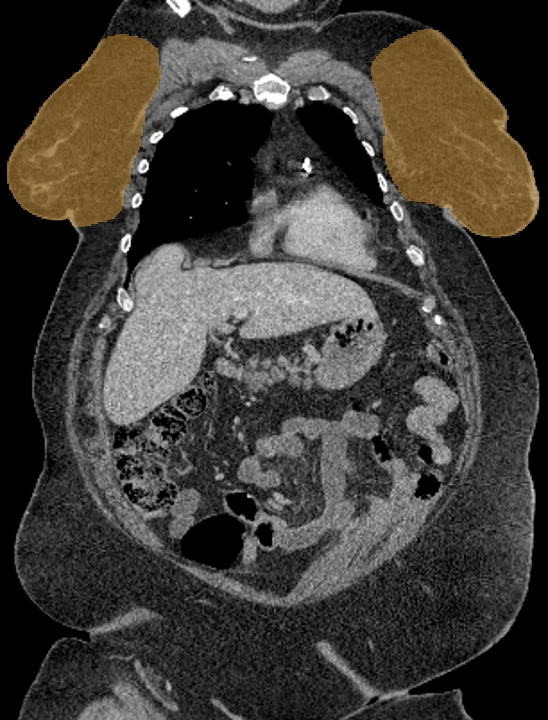
Breast tissue on CT
Breast tissue on chest CT scans gets one shared outline from TotalSegmentator, covering both sides. Many chest CT scans are made for other reasons, so researchers can measure breast tissue volume and Hounsfield values across existing data sets and set them beside mammographic density. On PET or SPECT, breast uptake can be read and set against a reference region such as the liver.
Find answers to research questions like: How much breast tissue does a chest CT scan show? How closely does density on chest CT follow density on mammography? How does uptake in breast tissue differ between groups?
Run this task →
Run it online in your browser, with no coding.