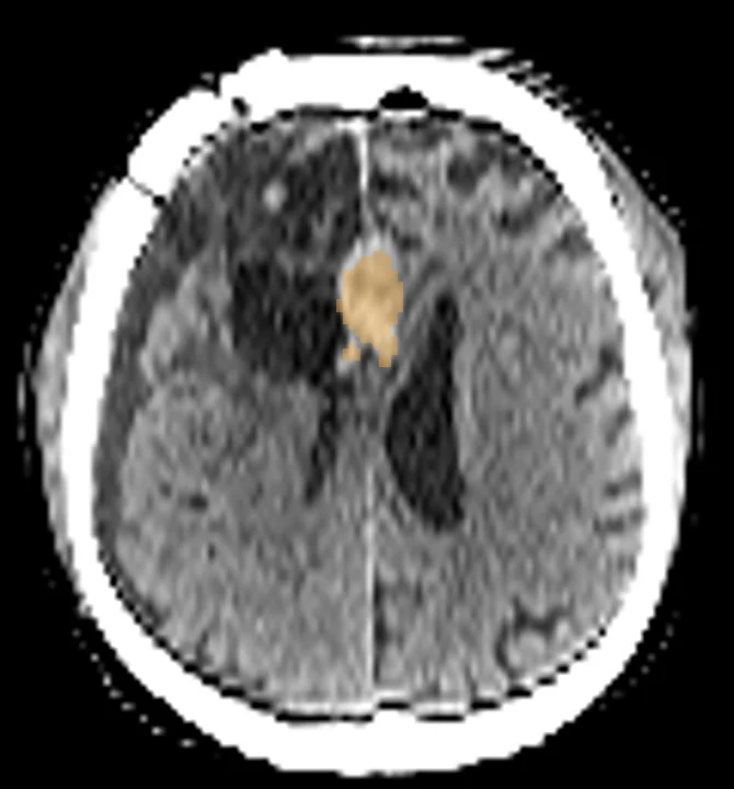
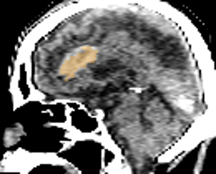
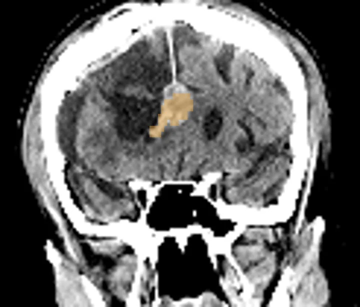
Cerebral bleeds on head CT
This TotalSegmentator AI task outlines intracerebral bleeds, also called intracerebral hemorrhage, on head CT scans. A study can then read bleed volume and Hounsfield values from the outline, set them beside the quick three-diameter estimate, and follow a bleed between a first and a later scan. An attached PET or SPECT scan adds uptake inside the outlined bleed.
Find answers to research questions like: How closely does outlined volume match the three-diameter estimate? How much does a bleed grow between two scans? How does uptake inside a bleed change between scan dates?
Run this task →
Run it online in your browser, with no coding.