Cerebral ventricle parts on CT
On a head CT scan, TotalSegmentator divides the lateral ventricles into frontal horn, occipital horn, temporal horn, body, and trigone on each side, and outlines the third and fourth ventricles. Measuring each part separately lets researchers compare groups, follow change between scans, and build reference values by age and sex. On PET or SPECT, these outlines read the uptake inside each ventricle and set it beside ventricle size.
Find answers to research questions like: Do the separate ventricle parts change by the same amount? How do ventricle volumes compare with a simple width index on the same scan? How does ventricle size relate to the uptake inside?
Run it online in your browser, with no coding.
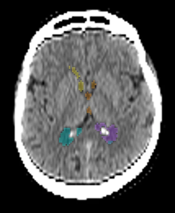
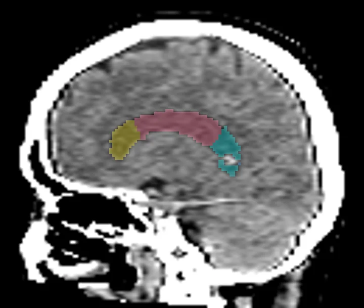
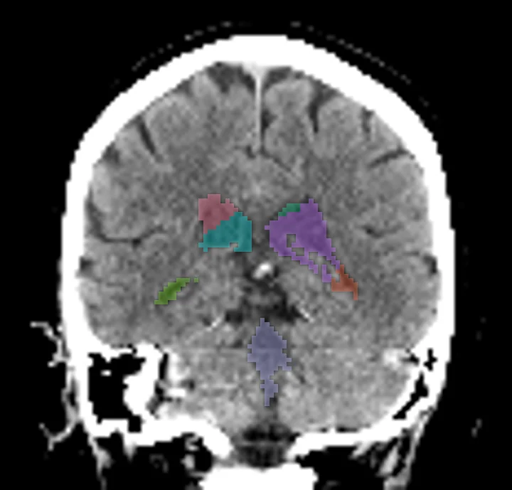
Found in this example
- Ventricle frontal horn left
- Ventricle occipital horn left
- Ventricle body left
- Ventricle temporal horn left
- Ventricle trigone left
- Ventricle frontal horn right
- Ventricle occipital horn right
- Ventricle body right
- Ventricle temporal horn right
- Ventricle trigone right
- Third ventricle
- Fourth ventricle
Example scan: head CT, TotalSegmentator dataset