Head glands and cavities on CT
Glands, cavities, and eye structures of the head are outlined on a CT scan: parotid and submandibular glands, nasal cavities, three levels of the pharynx, auditory canals, soft and hard palate, eyes, lenses, and optic nerves. TotalSegmentator gives research groups their volumes, to compare groups and follow change across repeated scans. On PET or SPECT, the same outlines read uptake inside each gland, to compare groups and scan dates.
Find answers to research questions like: How much do parotid and submandibular gland volumes change between scans? How does uptake in the parotid and submandibular glands differ between groups? Does pharynx volume differ with jaw and facial shape?
Run it online in your browser, with no coding.
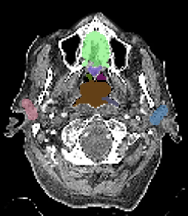
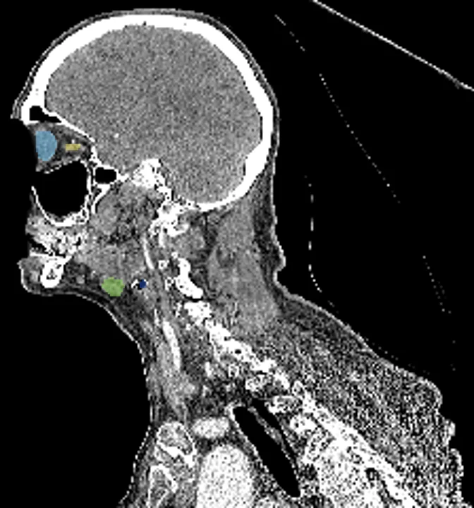
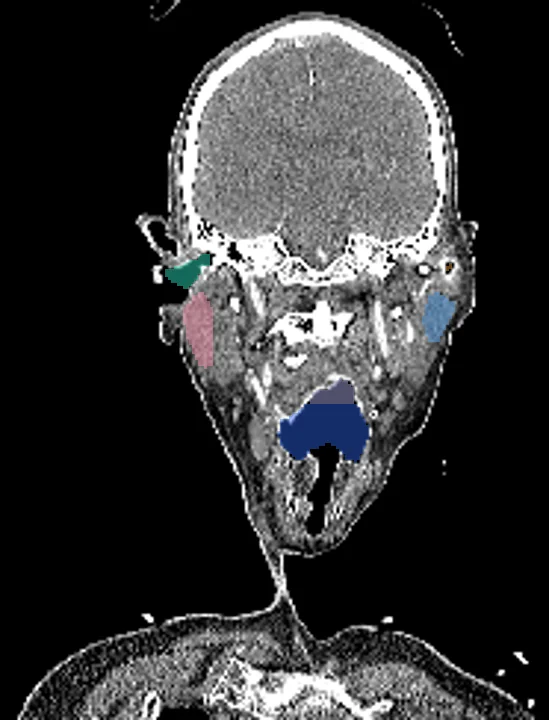
Found in this example
- Eye right
- Optic nerve right
- Parotid gland left
- Parotid gland right
- Submandibular gland right
- Nasopharynx
- Oropharynx
- Hypopharynx
- Nasal cavity right
- Nasal cavity left
- Auditory canal right
- Auditory canal left
- Soft palate
- Hard palate
- Eye left
- Optic nerve left
- Submandibular gland left
Also finds: Eye lens left, Eye lens right
Example scan: head CT, TotalSegmentator dataset