Head muscles on CT
The muscles of mastication are outlined per side on a head CT scan: masseter, temporalis, medial pterygoid, and lateral pterygoid, along with the digastric muscles and the tongue. With TotalSegmentator, studies can follow these volumes over time, relate them to age and body size, and test whether chewing muscle size reflects muscle mass elsewhere in the body. An attached PET or SPECT scan adds uptake per muscle.
Find answers to research questions like: Does masseter or temporalis size follow whole-body muscle mass? How much do these muscles change between two scans? How does uptake differ between the individual chewing muscles?
Run this task →
Run it online in your browser, with no coding.
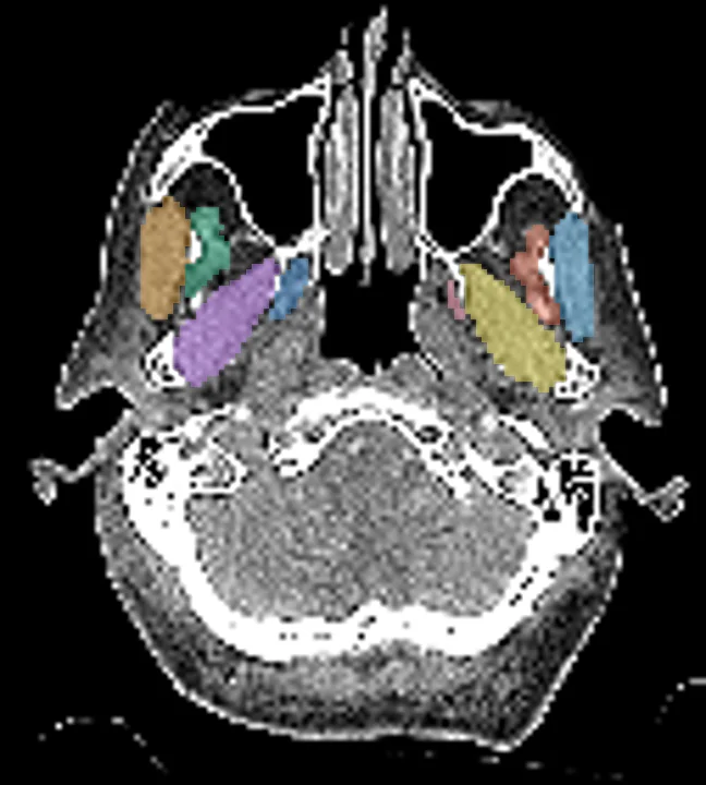
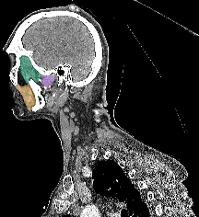
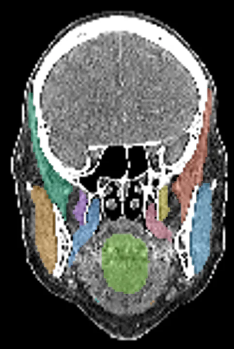
Found in this example
- Masseter right
- Masseter left
- Temporalis right
- Temporalis left
- Lateral pterygoid right
- Lateral pterygoid left
- Medial pterygoid right
- Medial pterygoid left
- Tongue
- Digastric right
- Digastric left
Example scan: head CT, TotalSegmentator dataset