Head and neck bones and vessels on CT
TotalSegmentator outlines bones, cartilage, and major vessels on a head and neck CT scan: the hyoid, zygomatic arches, styloid processes, thyroid and cricoid cartilages, air in the larynx, internal carotid arteries, and internal jugular veins. Research uses include reference values for anatomy, carotid size and length, and how these structures lie relative to each other. On PET or SPECT, uptake can be read along the carotid arteries, with the jugular veins as a blood-pool reference region.
Find answers to research questions like: How does carotid artery size vary with sex and body height? How do hyoid size and position change with age? How does carotid artery uptake compare with uptake in the jugular veins?
Run it online in your browser, with no coding.
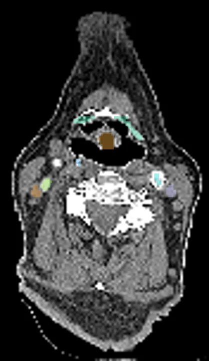
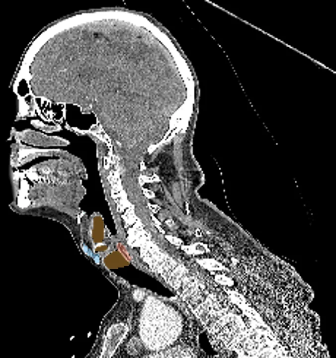
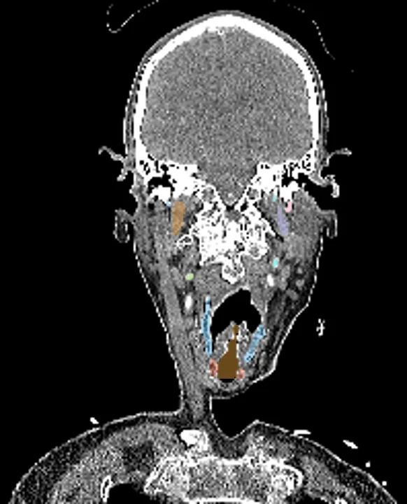
Found in this example
- Larynx air
- Thyroid cartilage
- Hyoid
- Cricoid cartilage
- Styloid process right
- Styloid process left
- Internal carotid artery right
- Internal carotid artery left
- Internal jugular vein right
- Internal jugular vein left
Also finds: Zygomatic arch right, Zygomatic arch left
Example scan: head and neck CT, TotalSegmentator dataset