Head and neck muscles on CT
A head and neck CT scan yields the sternocleidomastoid, trapezius, platysma, levator scapulae, three scalenes, sternothyroid, thyrohyoid, and prevertebral muscles on each side, plus the three pharyngeal constrictors. These TotalSegmentator outlines let studies compare neck muscle volume and CT density between groups and over time. An attached PET or SPECT scan adds uptake per neck muscle, to compare groups and scan dates.
Find answers to research questions like: Does neck muscle size follow muscle mass across the body? How do sternocleidomastoid, trapezius, and scalene volumes vary with age and sex? How does neck muscle uptake change between scan dates?
Run it online in your browser, with no coding.
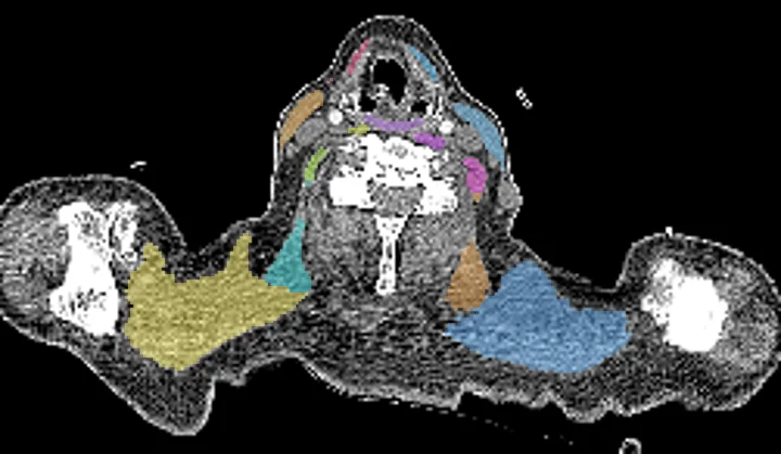
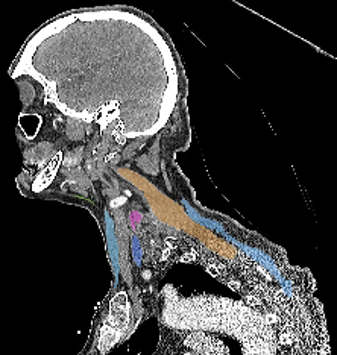
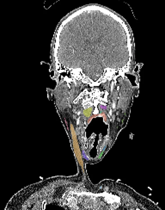
Found in this example
- Sternocleidomastoid right
- Sternocleidomastoid left
- Superior pharyngeal constrictor
- Middle pharyngeal constrictor
- Inferior pharyngeal constrictor
- Trapezius right
- Trapezius left
- Platysma right
- Platysma left
- Levator scapulae right
- Levator scapulae left
- Anterior scalene left
- Middle scalene right
- Middle scalene left
- Posterior scalene right
- Posterior scalene left
- Sterno thyroid right
- Sterno thyroid left
- Thyrohyoid right
- Thyrohyoid left
- Prevertebral right
- Prevertebral left
- Anterior scalene right
Example scan: head and neck CT, TotalSegmentator dataset