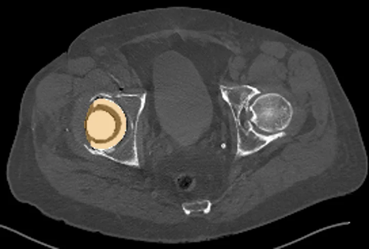
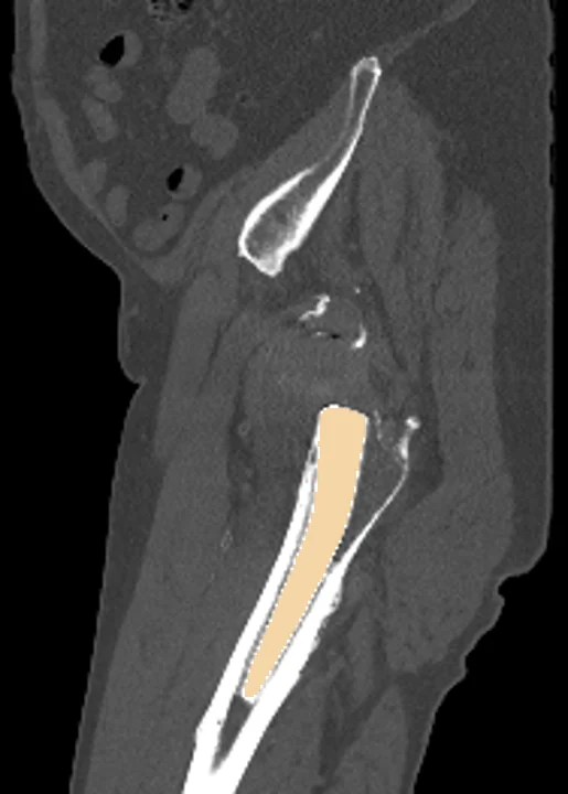
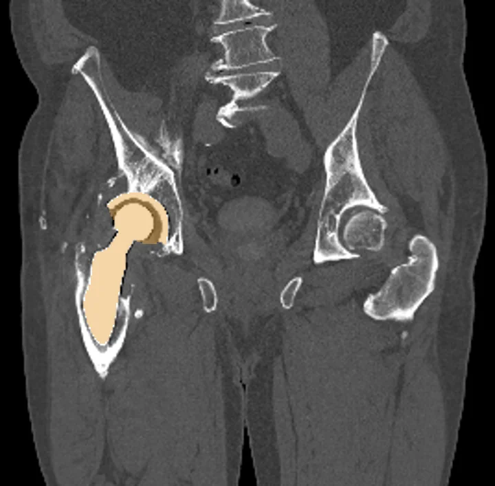
Hip implants on CT
TotalSegmentator outlines hip implants on pelvic CT scans and nothing else. The outline lets researchers measure where an implant sits, set it apart from nearby muscle and bone before measuring them, and study how metal artifacts affect the tissue around it. On a PET or SPECT overlay, the outline marks where the metal sits, which helps judge how it changes the values nearby.
Find answers to research questions like: How is the implant positioned in the pelvis across a study group? Does it move, or does the bone around it change, between two scans? How far from the metal do artifacts change uptake values?
Run this task →
Run it online in your browser, with no coding.