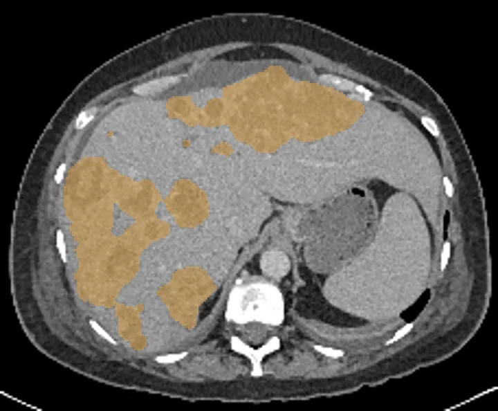
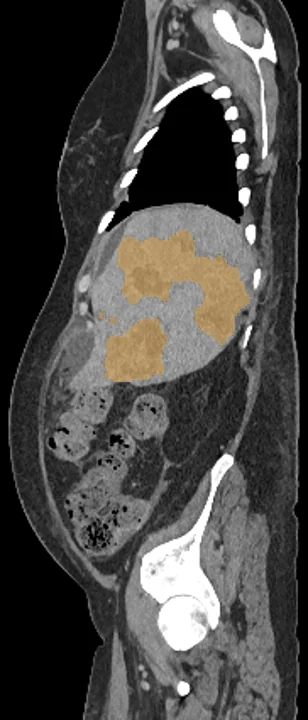
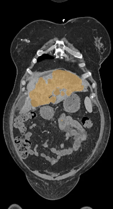
Liver lesions on CT
TotalSegmentator outlines every hepatic lesion on an abdominal CT scan as one shared structure. From that outline a study can measure total lesion volume, its share of the liver, and the Hounsfield values inside, then follow them across repeat scans. On PET or SPECT, lesion uptake can be read and set against the rest of the liver as a ratio, with lesion size taken into account.
Find answers to research questions like: What share of the liver do the lesions take up in different groups? How do lesion volume and CT density change between a baseline and a follow-up scan? Does lesion uptake relative to the surrounding liver differ between groups?
Run this task →
Run it online in your browser, with no coding.