Couinaud liver segments on CT
TotalSegmentator divides the liver on abdominal CT into Couinaud segments 1 to 8: the caudate lobe (1), left lateral (2 and 3), left medial (4), right anterior (5 and 8), and right posterior (6 and 7). CT volumetry of each segment lets researchers compare groups, follow change between scans, and place lesions by segment. On PET or SPECT, uptake can be read per segment, useful when the liver serves as a reference region.
Find answers to research questions like: How does each segment's share of the liver vary with age, sex, and body size? How do segment volumes change between a baseline and a follow-up scan? Which liver segment gives the steadiest reference uptake?
Run it online in your browser, with no coding.
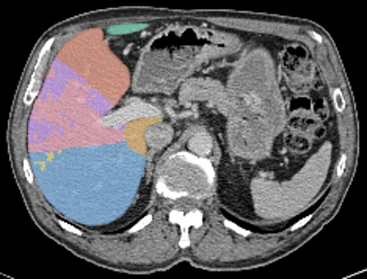
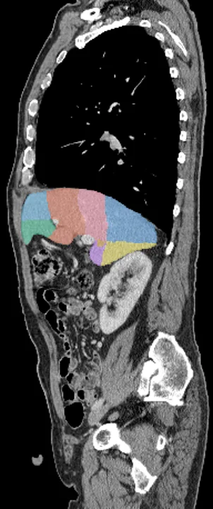
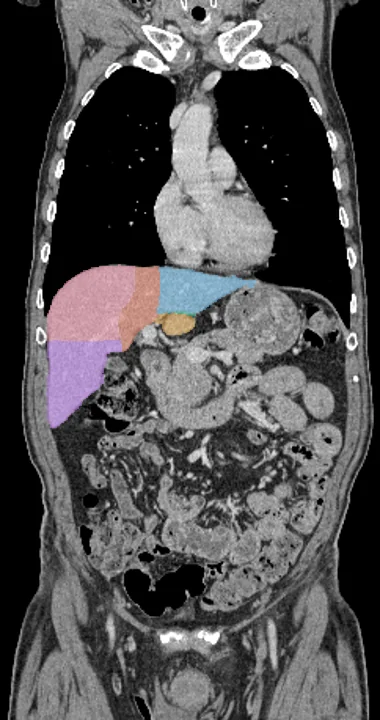
Found in this example
- Liver segment 1
- Liver segment 2
- Liver segment 3
- Liver segment 4
- Liver segment 5
- Liver segment 6
- Liver segment 7
- Liver segment 8
Example scan: chest to pelvis CT, TotalSegmentator dataset