Liver vessels and tumors on CT
TotalSegmentator outlines the hepatic vessels and liver tumors on abdominal CT scans, with one structure for all vessels and one for all tumors. Studies use them to measure vessel volume against liver size, follow tumor volume between scans, and ask how close tumors lie to the vessels. On PET or SPECT, tumor uptake can be read and set against the liver around it as a ratio.
Find answers to research questions like: How large is the vessel tree relative to the liver in different groups? How does total tumor volume change between two scans? How does tumor uptake compare with uptake in the surrounding liver?
Run this task →
Run it online in your browser, with no coding.
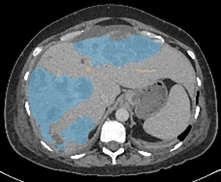
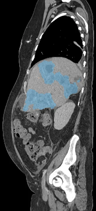
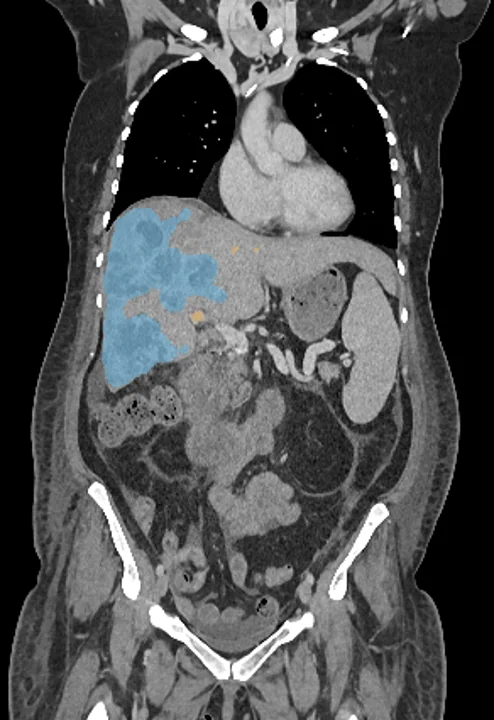
Found in this example
- Liver vessels
- Liver tumor
Example scan: chest to pelvis CT, TotalSegmentator dataset