Pleural and pericardial effusion on CT
Pleural effusion and pericardial effusion get separate outlines when TotalSegmentator segments a chest CT scan, and a third structure covers the lungs. Studies use them to measure fluid volume, follow effusions across repeat scans, and relate them to heart function and health outcomes. An attached PET or SPECT scan adds uptake inside the fluid, which can be compared between groups and scan dates.
Find answers to research questions like: How do quick estimates of pleural effusion volume compare with a full outline? How does uptake in the fluid change between scan dates? Is pericardial effusion volume linked to heart function?
Run this task →
Run it online in your browser, with no coding.
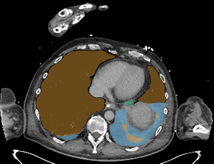
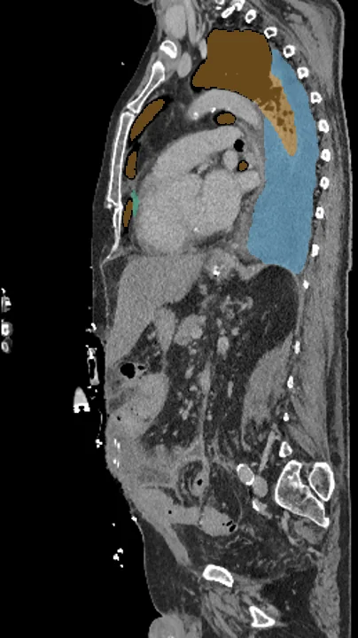
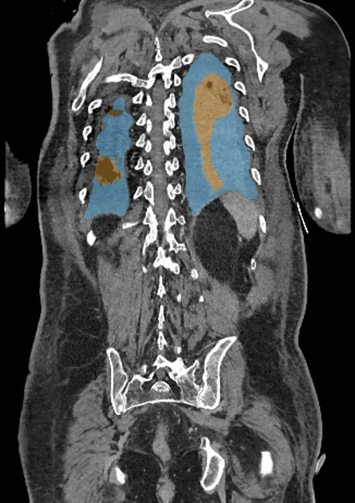
Found in this example
- Lung pleural
- Pleural effusion
- Pericardial effusion
Example scan: chest to pelvis CT, TotalSegmentator dataset