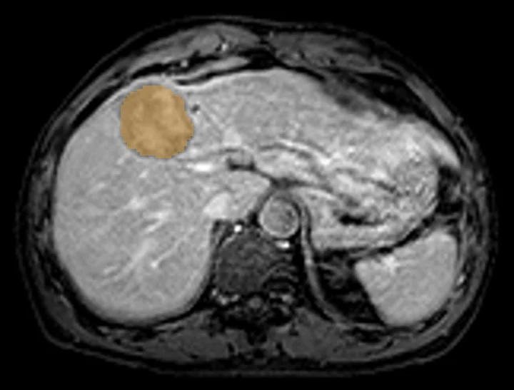
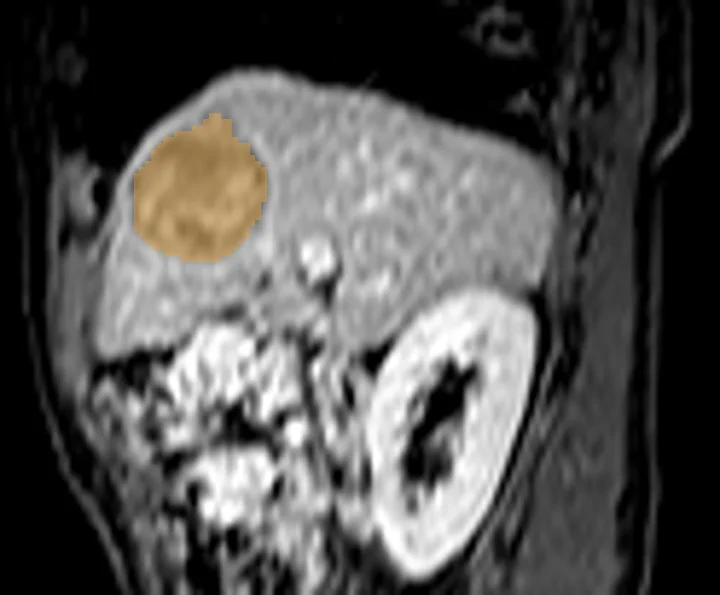
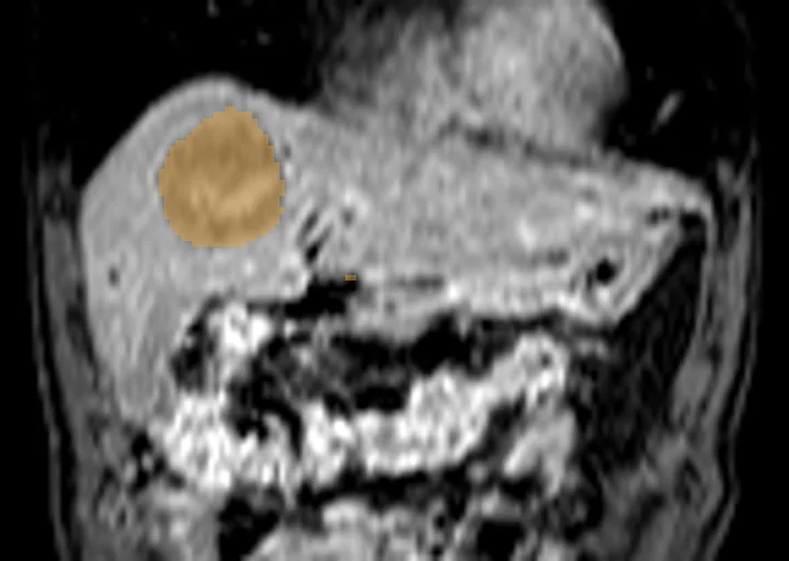
Liver lesions on MRI
TotalSegmentator MRI outlines all liver lesions on an MRI scan as one shared structure, a first draft to correct by hand before measuring. Studies use it for research outlining of lesion volume across repeated scans and to compare MRI with other scan types.
Find answers to research questions like: How large are the lesions on each MRI scan? How does total lesion volume change between a baseline and a follow-up MRI scan? Does lesion volume on MRI match lesion volume on CT in the same person?
Run this task →
Run it online in your browser, with no coding.