Bring AI to your
medical imaging research

Segment scans, generate synthetic images, and create research reports in your browser.

  • AI models for CT and MRI
  • No installation or coding required
  • Click-to-segment tools and manual editing
  • Track measurements across scans
  • Export results in standard formats
Browse models
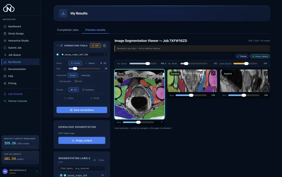
Product preview
Scroll
How it works

From scan to result in four steps

For tasks that use a scan; synthetic image generation starts without one.

Upload your medical image

Choose a file or folder from your device.

Pick an AI model from our library

See the cost before you run.

Submit & monitor AI tasks

Your task runs in the cloud.

View, adapt, and download results

Review in your browser. Export in standard formats.

Capabilities

Segment, generate, report — one platform

Image segmentation

Interactive and automatic segmentation

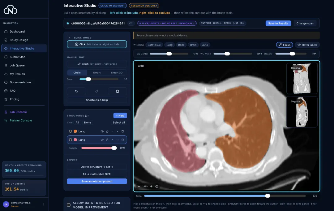
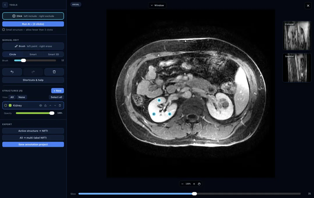
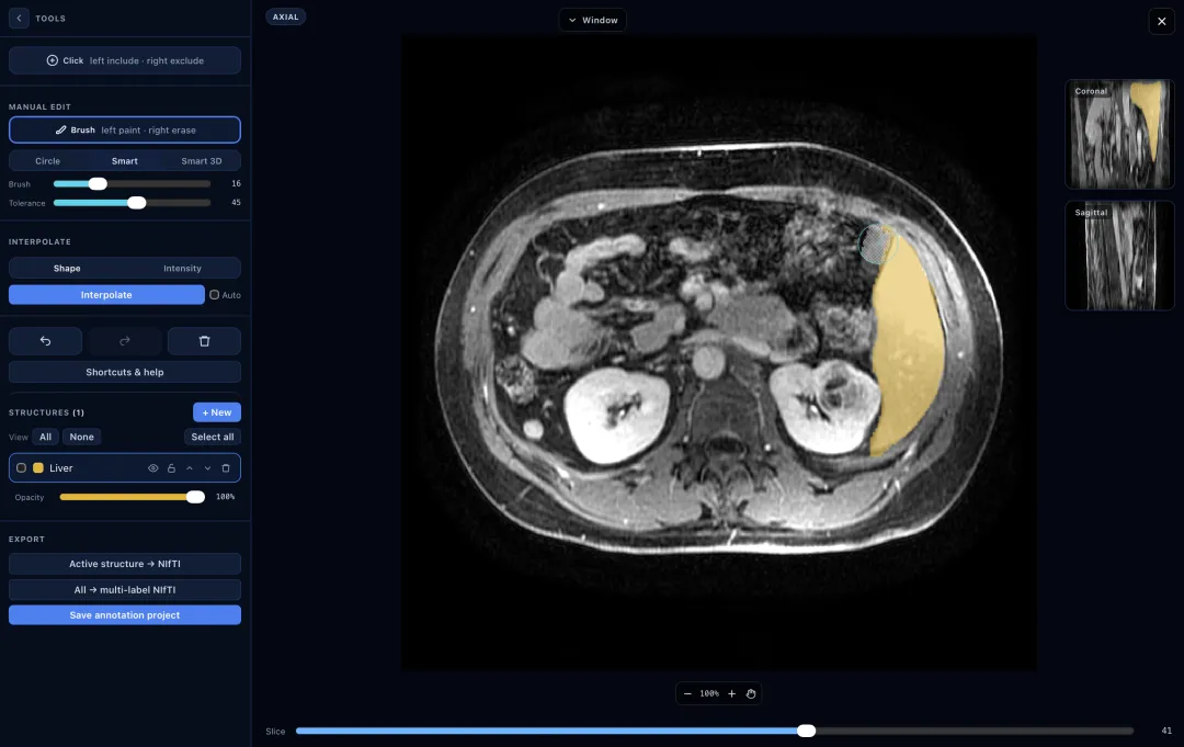
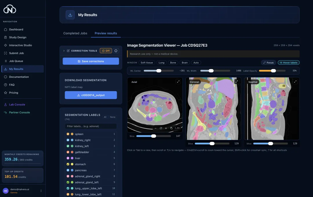
Segment CT and MRI scans automatically or with interactive clicks. Review and edit both in your browser.

Nalvera iSeg · in-house TotalSegmentator MOOSE MuscleMap CT / MRI
A brain MR with the cerebellum outlined in orange by the click-guided AI.
Output · brain MR · cerebellum
Image generation

Synthetic image generation

Generate synthetic CT or brain MRI images without an input scan, for research datasets, software testing, and demonstrations.

MAISI-v2 No input scan needed
A synthetic thorax CT slice, generated by AI and shown at lung window settings.
Output · thorax 3D CT
A synthetic T2-weighted brain MR slice, generated by AI.
Output · brain T2 MR
Report generation

Research reports from CT

Generate structured research reports from abdominal CT scans with Merlin.

Merlin · vision-language AI 13 organ systems
Input scan
The input scan for the research report example: an axial CT slice through the mid-abdomen showing both kidneys, the aorta, the vertebral body and gas-filled bowel loops, at soft-tissue window settings. Abdominal 3D CT
AI-generated report
[kidneys] The kidneys enhance homogeneously. No focal renal lesions. No hydronephrosis or hydroureter involving either collecting system. [bowel] There is no evidence of bowel obstruction. [circulatory] Atherosclerosis without aneurysm. [musculoskeletal] No suspicious osseous lesions identified. [peritoneum] No free fluid. Abdominal wall: normal. [lower thorax] No significant pulmonary, pleural, or mediastinal abnormality is seen in the visualized portions of the thorax. [liver] Normal in size. There are multiple subcentimeter hypodensities throughout both lobes of the liver, which likely represent cysts or hamartomas but are too small to characterize on this examination. No intrahepatic or extra hepatic ductal dilatation. [gallbladder] Surgically absent. [pelvic] Normal. Prostate and seminal vesicles: the prostate is mildly enlarged measuring 5 cm in transverse dimension (3/401). [lymph nodes] No enlarged retroperitoneal, mesenteric, pelvic or inguinal lymph nodes. [spleen] Normal. [pancreas] Normal. [adrenal glands] Normal.

Run your first AI task

Free to start. No credit card needed.